Body Fluid Biomarkers

                   Brain imaging including magnetic resonance imaging (MRI) has helped revolutionize our understanding of structural injury in neurological disease including the field of MS research.  Although brain imaging methods with some specificity for type of injury have been developed, methods for tracking the type of pathology and the specific cell types that manifest injury has been limited.  We aim to use biomarkers derived from body fluids (including plasma, serum and CSF) to help monitor neurological diseases especially degenerative, progressive diseases of the central nervous system. Using an understanding of the biology of MS as well as other neurodegenerative diseases of the CNS we aim to select and identify promising candidate biomarkers for detection and assay optimization. We then aim to refine methods for  the detection of possible CNS (and PNS) specific proteins and peptides including developing new tools for highly-quantifiable, highly-sensitive quantification of such proteins in blood for ready assessment and monitoring. Furthermore, we also aim to biologically validate these potential biomarkers using pre-clinical tools to document the source of these proteins and determine how such proteins and peptides get into the various compartments (blood, CSF, other body fluids) where they can be detected.  Ultimately we aim to use these approaches to improve our capacity to study CNS injury in both animal models and human diseases dynamically and to try and understand the interplay between various pathological processes that produce neurological dysfunction. 

This improved understanding will then be used to employ these improved biomarkers in ongoing and future clinical trials monitoring neuroprotective and neurorestorative therapies. The central aim of our projects is help monitor, understand and prevent irreversible damage to nerve cells and the nervous system. 

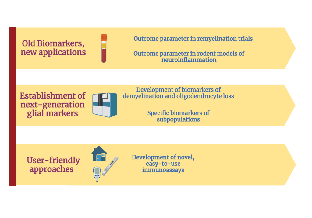
In the context of MS:  the ability to untangle the complexity of the various mechanisms that contribute to injury and to measure the effect of each of them independently is very limited. Therefore, our work focuses on the development, validation, and utilization of novel highly-sensitive technologies and state-of-the-art protein measurement and biomarker discovery tools to determine the protein signature in people with MS (pwMS) to:

  • Understand the mechanisms that ultimately cause neurodegeneration and irreversible disability.
  • Define the causes, and characterize the consequences of remyelination failure
  • Develop novel tools that allow for the wide-spectrum application of biomarkers in different clincal and research settings. 


Meet our national and international Collaborators:


Biomarker Background and Applications:

Biomarkers are measurable biological indicators that provide information about normal biological processes, pathogenic processes, or responses to interventions or environmental exposures. They can be molecules, genes, proteins, cells, or other measurable entities found in bodily fluids (such as blood, urine, or cerebrospinal fluid) or tissues. Biomarkers can be used for various purposes, including disease diagnosis, prognosis, monitoring treatment response, and assessing disease progression.


In the context of medicine and healthcare, biomarkers play a crucial role in improving the understanding, detection, and management of diseases. Here are some examples:

  1. Diagnostic biomarkers: These biomarkers can aid in the identification of a particular disease or condition. For instance, elevated levels of prostate-specific antigen (PSA) in blood samples are used as a diagnostic biomarker for prostate cancer.
  2. Prognostic biomarkers: These biomarkers provide information about the likely outcome or progression of a disease. They help predict how a disease may develop or the likelihood of specific treatment outcomes. For example, certain genetic markers are associated with a higher risk of developing certain types of cancer.
  3. Predictive biomarkers: These biomarkers help predict an individual's response to a specific treatment or intervention. They can guide healthcare professionals in choosing the most appropriate treatment strategy for a patient. For instance, the presence of specific genetic mutations in lung cancer can predict the response to targeted therapies.
  4. Monitoring biomarkers: These biomarkers are used to assess the effectiveness of a treatment or intervention and to monitor disease progression or recurrence. Blood tests measuring levels of specific proteins or tumor markers can be used as monitoring biomarkers in cancer patients.


Biomarkers are actively researched and utilized in various fields, including oncology, cardiology, neurology, infectious diseases, and many others. They have the potential to enhance early detection, personalized treatment plans, and improve patient outcomes by enabling more precise and targeted approaches to healthcare.

Biomarkers are valuable in multiple sclerosis (MS) research and clinical practice. They can help in the diagnosis, prognosis, monitoring of disease activity, and assessment of treatment response. Here are some examples of biomarkers used in MS:

  1. Magnetic Resonance Imaging (MRI) Biomarkers: MRI is a commonly used imaging technique in MS. Various MRI measures serve as biomarkers to assess disease activity and progression. These include the number and volume of brain lesions, the presence of contrast-enhancing lesions indicating active inflammation, and brain atrophy (loss of brain tissue volume) over time. These MRI biomarkers assist in diagnosis, monitoring disease activity, and evaluating treatment response.
  2. Cerebrospinal Fluid (CSF) Biomarkers: Analysis of CSF obtained through a lumbar puncture can provide valuable information in MS. Biomarkers such as oligoclonal bands (abnormal immune response markers) and specific proteins like immunoglobulin G (IgG) can help confirm the diagnosis and differentiate MS from other conditions. Additionally, specific CSF biomarkers may indicate disease activity or be associated with disease severity.
  3. Blood Biomarkers: Blood-based biomarkers are being investigated in MS to aid in diagnosis, prognosis, and treatment response assessment. Examples include serum antibodies (such as anti-myelin antibodies), cytokines, chemokines, and specific immune cells. These biomarkers have the potential to provide insights into disease mechanisms, identify disease subtypes, and predict treatment responses.
  4. Genetic Biomarkers: Genetic biomarkers associated with MS susceptibility and disease progression are being studied. Specific genetic variants, such as those in the human leukocyte antigen (HLA) region, have been linked to increased MS risk. Genetic biomarkers can help in identifying individuals at higher risk of developing MS and provide insights into disease mechanisms.


It's important to note that while biomarker research in MS advances, many biomarkers are still being studied and validated. Developing reliable and clinically meaningful biomarkers remains an ongoing area of research, aiming to improve early diagnosis, individualize treatment approaches, and predict disease course and treatment outcomes in MS.

Astrocyte biomarkers are used in multiple sclerosis (MS) research to gain insights into the underlying pathology, disease progression, and treatment responses. Astrocytes are a type of glial cell found in the central nervous system (CNS), including the brain and spinal cord. They play a crucial role in supporting neuronal function and maintaining the integrity of the CNS.

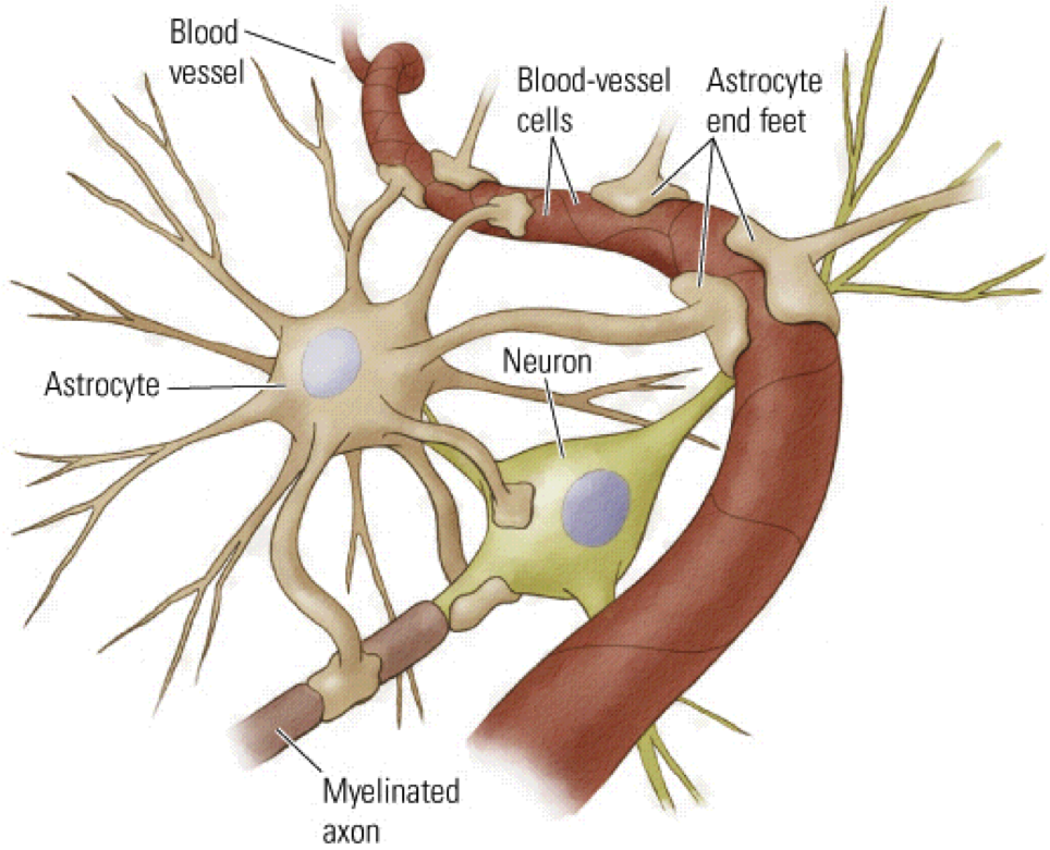
Here are some reasons why astrocyte biomarkers are used in MS research:

  1. Disease Pathology: Astrocytes are actively involved in the immune response and tissue repair processes in MS. They become activated in response to inflammation and release various molecules that can contribute to neuroprotection or damage. By studying astrocyte biomarkers, researchers aim to understand the specific functions and mechanisms astrocytes contribute to MS pathology.
  2. Neuroinflammation and Disease Activity: Astrocytes play a role in neuroinflammation, an essential process in MS. They can release proinflammatory molecules and contribute to forming the blood-brain barrier (BBB) breakdown. Astrocyte biomarkers, such as glial fibrillary acidic protein (GFAP) and S100B protein, are studied to assess disease activity and neuroinflammatory processes in MS.
  3. Disease Progression and Neurodegeneration: In MS, neurodegeneration occurs gradually and contributes to the development of disability over time. Astrocytes are involved in supporting neuronal health and synapse function. Dysfunction of astrocytes can lead to neurodegenerative processes. By identifying astrocyte biomarkers, researchers aim to understand the contribution of astrocyte dysfunction to disease progression and neurodegeneration in MS.
  4. Treatment Responses: Astrocytes may be involved in responding to MS treatments, including disease-modifying therapies (DMTs). Biomarkers associated with astrocyte activity can provide insights into treatment effects on astrocyte function, inflammation, and neuroprotection. Monitoring astrocyte biomarkers may help evaluate treatment responses and assess the efficacy of different therapeutic approaches in MS.
  5. Therapeutic Targets: Identifying astrocyte-specific biomarkers can aid in the development of new therapeutic targets for MS. Understanding the specific molecular pathways and signaling mechanisms in astrocytes can potentially lead to the development of novel treatment strategies aimed at modulating astrocyte function and neuroinflammation.

It's important to note that astrocyte biomarker research is still ongoing, and further studies are needed to validate and establish their clinical utility in MS diagnosis, prognosis, and treatment. However, the investigation of astrocyte biomarkers holds promise in advancing our understanding of MS pathophysiology and improving therapeutic approaches in the future.


In the Biomarker work that we focus on, we utilize different immunoassays for the early detection of MS. 

Our ELISA machine

ELISA stands for Enzyme-Linked Immunosorbent Assay. It is a laboratory technique used to detect and quantify the presence of specific proteins, antibodies, or other molecules in biological samples. ELISA is widely used in various research fields, clinical diagnostics, and biotechnology.

Here's a general overview of how ELISA works:

  1. Coating: The first step involves coating a solid surface, such as a microplate well, with a capture antibody. The capture antibody is specific to the molecule of interest and can bind to it.
  2. Blocking: After coating, the plate is typically treated with a blocking agent, such as bovine serum albumin (BSA), to prevent nonspecific binding of other molecules to the surface.
  3. Sample and Standards: The sample (e.g., serum, plasma, cell lysate) or standards with known concentrations of the molecule of interest are added to the coated wells. If the molecule is present in the sample, it will bind to the capture antibody.
  4. Detection: A detection antibody is then added to the wells. This antibody recognizes a different epitope on the molecule of interest and is conjugated to an enzyme, such as horseradish peroxidase (HRP) or alkaline phosphatase (AP). The detection antibody binds to the captured molecule, forming a sandwich-like complex.
  5. Substrate Addition: The addition of a substrate-specific to the enzyme conjugated to the detection antibody leads to a reaction that produces a measurable signal. The substrate is converted by the enzyme into a detectable product, such as a colored or fluorescent compound.
  6. Signal Measurement: The intensity of the generated signal is proportional to the amount of the molecule of interest present in the sample. This signal can be measured using a spectrophotometer or a plate reader, which provides quantitative data.

ELISA allows for the quantification of proteins, antibodies, hormones, cytokines, and other molecules in various samples, such as blood, serum, plasma, urine, or cell lysates. It offers high sensitivity and specificity, making it a valuable tool in research, clinical diagnostics, and drug discovery.

There are different variations of ELISA, including direct, indirect, sandwich, competitive, and multiplex ELISAs, each designed to suit specific experimental requirements and target molecules.


Direct ELISA:

In a direct ELISA, the target antigen (or molecule of interest) is directly immobilized onto the solid surface (e.g., microplate well) and detected using a labeled primary antibody specific to the antigen. The primary antibody is conjugated to an enzyme, such as horseradish peroxidase (HRP) or alkaline phosphatase (AP), which generates a detectable signal when a substrate is added.

Direct ELISAs are relatively simple and have fewer steps, but they require the availability of a high-quality specific primary antibody.


Indirect ELISA:

In an indirect ELISA, the target antigen is coated onto a solid surface, similar to the direct ELISA. Instead of using a labeled primary antibody, an unlabeled primary antibody specific to the antigen is added. A secondary antibody, which is conjugated to an enzyme, is then added. The secondary antibody recognizes and binds to the primary antibody.

The advantage of an indirect ELISA is that a single labeled secondary antibody can be used with multiple primary antibodies, making it more flexible and cost-effective.

Indirect ELISAs are commonly used when high sensitivity is required or when primary antibodies are available against the target antigen.

Figure 1: Indirect ELISA Steps. The Clear Polystyrene Microplates that the ELISA takes place in have a strong enough bond to the protein that it sticks after incubation for 1 hr. The Block Step is important to make sure that there is not too much background (non-specific binding or signal that may be observed in the absence of the specific target molecule. It represents any signal or noise that is unrelated to the presence of the target analyte.) Wash steps are important to wash away any residual reagents; however, do not leave them on too long or it will wash away your work/ inversely wash too little and you’ll have a high background. The strongest bond is the streptavidin to HRP and is extremely stable. Adding the primary and secondary antibodies is important to house the streptavidin-HRP bond. The HRP also allows us to have color for quantification in the machine. 


Sandwich ELISA:

Sandwich ELISA is used to detect and quantify antigens that have multiple epitopes, allowing for the formation of a "sandwich" complex. The solid surface is coated with a capture antibody specific to the target antigen. The antigen in the sample is then added, and it binds to the capture antibody. A detection antibody, which is specific to a different epitope on the antigen, is added, usually labeled with an enzyme.

The sandwich complex is formed when the detection antibody binds to the captured antigen.

This technique provides high specificity and sensitivity, as the antigen is captured between two antibodies, enabling detection even when the antigen is present in low concentrations.

Figure 2: Sandwich ELISA steps. The capture antibody MUST incubate overnight to create a strong bond to the well plate. Again the block is important so we do NOT have too much background. Step 10 for the biotinylated linker is only used when we do NOT have a biotinylated detection antibody. However we do have biotinylated antibodies for this ELISA. 


Competitive ELISA:

In a competitive ELISA, the sample antigen competes with a labeled antigen (conjugated to an enzyme) for binding to a limited amount of specific antibodies. The solid surface is coated with the capture antibody. A mixture of the labeled antigen and the sample antigen is added to the well, allowing them to compete for binding to the capture antibody. The amount of labeled antigen bound to the capture antibody is inversely proportional to the concentration of the sample antigen.

Competitive ELISAs are useful when measuring the presence or concentration of small molecules, such as hormones or drugs, in a sample. 


Each type of ELISA has its advantages and limitations, and the choice of which type to use depends on the specific experimental requirements, target molecule, and available reagents. Understanding the differences between these ELISA variations helps researchers select the most appropriate assay design for their specific applications.

Overall, ELISA is a versatile and widely used technique for detecting and quantifying specific molecules in biological samples, contributing to advancements in various fields of science and medicine.


Our MSD machine

Meso Scale Discovery (MSD) platform is a technology that combines electrochemiluminescence with multi-spot microplate technology to enable highly sensitive and precise measurement of biomarkers in biological samples.

The MSD assay offers several advantages over traditional ELISA (enzyme-linked immunosorbent assay) methods.

Here are some key features of the MSD assay:

  1. Sensitivity: The MSD platform provides enhanced sensitivity, allowing the detection of low-abundance biomarkers in small sample volumes. This is particularly useful when measuring biomarkers present at low concentrations.
  2. Wide Dynamic Range: The MSD assay has a wide dynamic range, enabling accurate quantification of both high and low concentrations of analytes in the same sample. This flexibility is advantageous when analyzing samples with varying biomarker levels.
  3. Multiplexing Capability: The MSD platform allows for multiplexing, which means it can simultaneously measure multiple analytes in a single sample. This feature saves time, reduces sample consumption, and enables the study of multiple biomarkers simultaneously. 
  4. Precision and Reproducibility: The MSD assay demonstrates high precision and reproducibility, minimizing variability and ensuring reliable and consistent results across experiments.
  5. Customizable Panels: MSD offers pre-configured assay panels targeting specific biomarkers or disease pathways. Additionally, custom panels can be created to measure analytes of interest based on specific research needs.

The workflow of an MSD assay involves the immobilization of capture antibodies on specific spots within the microplate wells. After adding the sample and appropriate detection antibodies labeled with electrochemiluminescent tags, electrochemical stimulation induces the release of light proportional to the bound analyte. The emitted light is then measured and quantified by the MSD instrument.

MSD assays have found applications in various fields, including drug discovery, clinical research, biomarker validation, and translational medicine. They are particularly useful when analyzing complex biological samples, such as serum, plasma, cell lysates, or tissue extracts, for the quantitative measurement of proteins, cytokines, growth factors, and other biomolecules.

It's worth noting that the MSD platform and associated assays are proprietary to Meso Scale Discovery and represent a specific technology within the broader field of immunoassays.


Our SIMOA machine

SIMOA stands for Single Molecule Array, which is an ultra-sensitive immunoassay technology developed by Quanterix Corporation. SIMOA is designed to detect and quantify low levels of biomarkers or analytes with exceptional sensitivity and precision.

The SIMOA technology utilizes a digital ELISA approach, where individual target molecules are captured and detected as discrete units, or "digital" signals, rather than measuring the average signal of a population of molecules. This allows for the detection and quantification of extremely low concentrations of analytes in biological samples.

The workflow of a SIMOA assay involves the following steps:

  1. Microbead Capture: Microbeads with capture antibodies specific to the target analyte are used to capture the molecules of interest from the sample.
  2. Wash: Unbound components are removed through washing steps to minimize background noise.
  3. Detection: Detection antibodies labeled with unique DNA oligonucleotide tags are introduced. If the target analyte is present, the detection antibody binds to it, forming an antibody-antigen complex.
  4. Digital Barcoding: Each captured bead is placed into an array of femtoliter-sized wells, called an array of microwells, or "nanowells." These nanowells can only accommodate one bead each, allowing for the isolation and counting of individual captured analyte molecules.
  5. Amplification and Imaging: In the nanowells, DNA amplification occurs to generate many copies of the DNA tags associated with the captured analyte molecules. The amplified DNA tags are then labeled with fluorescent probes and imaged using a specialized imaging system.
  6. Data Analysis: The imaging data is analyzed, and the number of positive signals or "digital" events is counted. This event counting allows for the quantification of the target analyte in the original sample.

SIMOA technology offers significant advantages in terms of sensitivity, dynamic range, and precision compared to conventional immunoassays. It has been employed in various areas of research, including biomarker discovery, diagnostics, and therapeutic development, to detect and monitor analytes at extremely low concentrations.

It's worth noting that the term SIMOA specifically refers to the technology developed by Quanterix Corporation. Other companies and research institutions may have developed similar ultra-sensitive immunoassay technologies with different names or variations.


We are currently using our SIMOA runs with EPIC samples (human samples of serum at different timepoints). EPIC (Expression, Proteomics, Imaging, Clinical) is an intensive observational study of over 500 people with Multiple Sclerosis (MS) who have been carefully studied since 2004. EPIC employs the most advanced imaging, molecular, cellular and bioinformatics techniques to gain greater understanding of MS susceptibility and long-term progression.